Life, at its core, is built from a single, microscopic building block. It’s the smallest anatomical unit capable of independent existence. Every organism you can name—from the towering redwood to the invisible bacteria in your gut—is made of these components. They have a job description that hasn’t changed in billions of years: eat, interact with the environment, and reproduce.
But not all cells are created equal. The biological world splits them into two distinct camps. On one side, you have eukaryotic cells. These are the complex workers found in plants, animals, fungi, and protists. They have a command center. On the other side are prokaryotic cells. Simpler. Older. They lack that central command center. This distinction matters because it dictates how an organism functions, grows, and survives.
The Basic Anatomy of a Cell
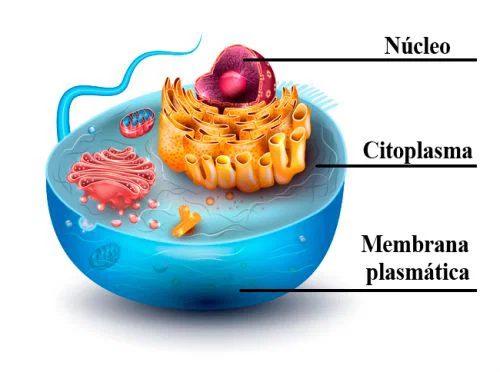
Most cells, regardless of their complexity, share three fundamental parts. Think of them as the non-negotiable requirements for being alive.
First, there’s the plasma membrane. This isn’t just a wall. It’s a selective barrier. It controls what goes in and what stays out, maintaining the delicate chemical balance required for life. Without it, the cell dissolves into the surrounding medium.
Second is the cytoplasm. This is the gel-like substance filling the cell. It’s where the magic happens. Chemical reactions occur here. Proteins are synthesized. It’s the stage upon which cellular life plays out.
The third part depends on the cell type. In eukaryotic cells, there is the nucleus. This is the brain of the operation. It houses the DNA, the genetic blueprint. It directs protein synthesis and cellular reproduction.
Prokaryotic cells lack a nucleus. Their DNA floats freely in the cytoplasm. This structural difference is the primary reason eukaryotes can achieve much greater complexity and size.
Beyond the Basics: Support and Structure
If you look closer, the picture gets more detailed. The three basic parts don’t work alone. They rely on supporting structures that maintain shape and internal transport.
Inside the cytoplasm lies the cytoskeleton. It’s not a skeleton in the bony sense. It’s a network of protein filaments. It provides structural support. It allows the cell to change shape. It helps move organelles around like a tiny logistics network.
Then there are the organelles. These are specialized structures within the cytoplasm, each with a specific function. Mitochondria generate energy. Ribosomes build proteins. They are the organs of the cell, working in concert to keep the organism alive.
Finally, some cells have a cell wall. You won’t find this in human cells. It’s present in plants, fungi, and bacteria. It sits outside the plasma membrane. It provides rigid protection and structural support, allowing plants to stand upright against gravity.
The diversity of life stems from how these simple components are arranged and combined. A neuron and a skin cell look and act differently because they express different parts of the same basic cellular toolkit. Understanding this foundation is the first step to understanding biology itself.
The Nucleus: The Command Center of the Eukaryotic Cell
If you’ve ever wondered what keeps a cell from descending into chaos, look no further than the nucleus. It’s the big, central boss in eukaryotic cells. Prokaryotes don’t get this luxury. They just float their DNA around in the cytoplasm like it’s a public park. But eukaryotes? They build walls.
The nucleus is essentially a vault. It’s wrapped in a double membrane (the nuclear envelope) that separates the genetic treasure trove from the messy business of the cytoplasm. This isn’t just for show. The membrane controls what gets in and out, acting as a bouncer for the cell’s most important data.
Why the Nucleus Matters in Everyday Life
You might think of the nucleus as just a blob inside a cell. But it’s actually the reason you exist as a distinct individual. It stores the blueprint. Without it, there’s no instruction manual for building proteins. No instruction manual means no muscles. No heart. No brain. The nucleus coordinates the entire operation. It tells the ribosomes what to build and when to stop. It’s not just storage; it’s management.
How the Nucleus Coordinates Cellular Reproduction
Ever wonder how a single cell becomes two identical cells during healing or growth? The nucleus handles the heavy lifting here. Before a cell divides, the nucleus replicates its DNA. It makes exact copies of the genetic code so that each new cell gets a full set of instructions. This process is tightly regulated. If the nucleus messes up the replication, you get mutations. Cancer is often the result of the nucleus losing control over this reproduction process.
Structure of the Nucleus: More Than Just a Bag
The nucleus isn’t an empty room. It’s packed with machinery. Inside, you find chromatin — the DNA wrapped around proteins. When the cell is preparing to divide, this chromatin condenses into visible chromosomes. But when the cell is resting, it’s looser. Why does this matter? It affects how genes are expressed. Tight packing silences genes. Loose packing allows them to be read.
The nucleus is not just a repository for DNA; it is the active control room that dictates cellular behavior through gene expression.
Key Functions Beyond Storage
While storing genetic information is the headline act, the nucleus does other critical work.
- Gene Expression Regulation: It decides which genes get turned on or off based on the cell’s needs. A skin cell and a liver cell have the same DNA, but the nucleus uses different parts of it for each.
- Ribosome Production: The nucleus contains the nucleolus. This is where ribosomes are assembled. Ribosomes are the protein factories scattered throughout the cytoplasm. No nucleolus means no ribosomes. No ribosomes means no protein synthesis. The cell dies.
- Coordination of Organelle Work: The nucleus sends out signals and mRNA instructions that direct the mitochondria, endoplasmic reticulum, and Golgi apparatus. It’s the conductor of the cellular orchestra.
Where the Nucleus Fits in the Cell
The nucleus is suspended in the cytoplasm. It doesn’t float freely. It’s tethered to the cytoskeleton, a network of protein filaments that gives the cell its
The Control Center and Its Contents
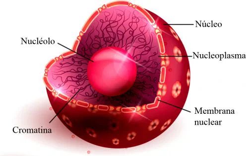
The nucleus isn’t just a static container. It’s a highly regulated facility wrapped in a double-layered membrane. This outer shell controls traffic, deciding what gets in and out. Inside, the environment is defined by nucleoplasma. Think of this as the internal fluid medium. It holds the structural components in suspension. Specifically, it cradles two key players: the nucleolus and chromatin.
The nucleolus operates on a strict schedule. Its primary job is ribosome production. It programs the formation of these cellular machines. Once assembled, ribosomes are transported out of the nucleus. They move into the cytoplasm to begin protein synthesis. Without this precise coordination, the cell loses its ability to build essential proteins.
Chromatin provides the genetic blueprint. It consists of DNA intertwined with proteins. When the cell prepares to divide, this loose structure condenses into chromosomes. This transformation ensures genetic material is packaged correctly for distribution. It’s a dynamic relationship. The chromatin exists to store information while remaining accessible for transcription.
The Cytoplasm: The Busy Workfloor
Beyond the nucleus lies the cytoplasm. It’s not just empty space filling the cell. It’s a bustling metabolic hub. The term refers to the entire region between the plasma membrane and the nuclear envelope. It includes the cytosol—the gel-like substance—and all the organelles suspended within it.
The cytosol itself is an aqueous solution. It contains water, salts, and organic molecules. Enzymes drift through this medium, catalyzing reactions. Metabolic pathways like glycolysis occur here. These processes break down glucose to produce energy. This energy powers everything the cell does.
Organelles anchor themselves in this matrix. Mitochondria generate ATP. The endoplasmic reticulum synthesizes proteins and lipids. The Golgi apparatus modifies and packages these products. Each structure has a specific location and function, yet they all interact. The cytoplasm facilitates this communication. It allows materials to diffuse or be actively transported between compartments.
Without the cytoplasm’s support, the nucleus would be isolated. The genetic instructions inside would have no means of execution. The ribosomes produced in the nucleolus need the cytoplasm’s resources to function. The chromatin’s code requires the cellular machinery embedded in this fluid environment.
The interplay between the nucleus and cytoplasm is constant. Signals travel both ways. Proteins made in the cytoplasm can enter the nucleus to regulate gene expression. This feedback loop maintains homeostasis. It allows the cell to adapt to changing conditions. Disruptions in this balance can lead to disease. Cancer, for instance, often involves failures in nuclear-cytoplasmic transport or gene regulation.
Understanding this division of labor explains how complex life functions. It’s not just about having parts. It’s about how those parts interact in a structured yet flexible environment. The nucleus holds the plan. The cytoplasm builds the structure.
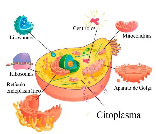
The cytoplasm is the jelly-like substance filling the inside of a cell. It is not just empty space. It is a busy, aqueous medium where life’s most basic chemical reactions happen. Two main components define it: the cytoskeleton and organelles.
In prokaryotic cells, which lack a nucleus, the cytoplasm holds the genetic material. This is a single molecule of DNA floating freely. The cytoplasm does the heavy lifting here. It keeps the genetic code safe and accessible.
The Cytoskeleton: The Cell’s Dynamic Frame
Think of the cytoskeleton as the cell’s internal scaffolding. It is not a rigid skeleton like yours. It changes constantly. It is a dynamic network of filaments present in every type of cell.
Its job is simple but critical. It gives the cytoplasm shape. It provides consistency. It also adds dynamism. Without it, the cell membrane would have no structure to support. The cytoplasm would be a shapeless blob. The cytoskeleton allows the cell to move, divide, and maintain its form against external pressures.
Organelles: The Tiny Factories
Organelles are small organs within the cytoplasm. They perform specific tasks. The variety and number of these organelles depend on the cell type. A muscle cell needs different tools than a leaf cell.
Here are the key players inside the cytoplasm:
Ribosomes : These are the only organelles found in all cells. They consist of two subunits made of ribosomal RNA (rRNA). Their function is protein synthesis. They build enzymes and other necessary proteins.
Endoplasmic Reticulum (ER) : This is a system of membranes that transports proteins. It has two parts. The rough ER receives proteins. The smooth ER creates new membranes.
Golgi Apparatus : These are sacs located near the nucleus. They process molecules transported by the ER. They package these molecules into small vesicles. These vesicles then release their contents outside the cell.
Lysosomes : These act as digestive bags. They have membranous walls that activate when needed. They digest material the cell ingests. Think of them as the cell’s waste management system.
Mitochondria : These are double-membrane sacs. They produce energy for cellular work through chemical reactions. Each mitochondrion has its own chromosome. It has its own DNA. This supports the theory that they were once independent organisms.
Vacuoles : These are compartments mainly in plant cells. They store fluids like water. They can also hold enzymes and nutrients.
Plastids : Found only in plant cells. They handle photosynthesis, starch storage, and synthesis of various materials. Chloroplasts, amyloplasts, and leucoplasts are common types.
Peroxisomes : Vesicle-shaped organelles. They oxidize and eliminate hydrogen peroxide. This protects the cell from toxic byproducts.
Centrioles : A pair of rod-shaped organelles that cross each other. They participate in cell division.
Flagella : A single protrusion from the cell. It helps with propulsion.
Cilia : Fine, multiple projections on the cell surface. They help cells move or move fluids across the cell.
The Plasma Membrane: The Gatekeeper
Every cell has a plasma membrane. It is also called the cell membrane or plasmalema. It is the boundary of the cell.
Function of the Plasma Membrane
- Keep the cell stable.
- Select which molecules enter or exit.
- Establish communication with other cells.
Structure of the Plasma Membrane
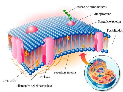
The cell membrane, with its fluid mosaic of lipids and proteins, is not the outermost boundary for every living thing. For plants, fungi, and many bacteria, there is another layer entirely. This is the cell wall.
It’s a structural shell. Rigid. Protective. And in many ways, more like a cage than a skin.
What is the Cell Wall?
While the plasma membrane controls what enters and leaves, the cell wall provides the shape. It prevents the cell from bursting under internal pressure. Without it, a plant cell in a hypotonic solution would simply swell and pop. The wall holds it together.
The composition varies wildly depending on who you ask—and who is being asked.
Cellulose dominates in plants. It’s a tough structural polysaccharide. Long chains of glucose linked together, forming microfibrils that act like steel rebar in concrete. This gives wood its strength and leaves their rigidity.
Chitin is the choice for fungi. You’ll find it in mushroom caps and insect exoskeletons. It’s nitrogen-containing. Strong. Flexible enough to allow growth but tough enough to resist degradation.
Bacteria use something else entirely. Peptidoglycan. A mesh of sugars and amino acids. It’s unique to prokaryotes. And it’s the target for antibiotics like penicillin, which disrupt its formation.
Why Does It Matter?
This isn’t just about structure. The wall is a barrier. A filter. A communication hub.
It prevents osmotic lysis. That’s the main job. But it also protects against mechanical stress. Think about a tree standing in a gale. The cells in its trunk are under constant tension. The wall bears that load.
It also plays a role in signaling. Plants detect pathogens not just through receptors in the membrane, but by changes in the wall’s integrity. When a bacterium breaks through, the plant senses the breach. It triggers a defense response. Rapid. Localized. Often fatal to the invader.
And let’s not forget growth. The wall isn’t static. Cells expand by loosening the cross-links in the peptidoglycan or cellulose matrix. Enzymes cut the chains. Turgor pressure pushes the membrane outward. The wall yields. Then it re-reinforces. It’s a dynamic process. Constant adjustment.
Differences Across Kingdoms
Not all walls are created equal. Here’s how they stack up:
- Plant cells : Primarily cellulose. Hemicellulose and pectin act as the glue. Lignin adds extra hardness in woody tissues.
- Fungal cells : Chitin. Sometimes glucans. No cellulose.
- Bacterial cells : Peptidoglycan. Gram-positive bacteria have thick layers. Gram-negative have thin layers plus an outer membrane.
- Archaea : Pseudopeptidoglycan or S-layers. No peptidoglycan. Different chemical bonds. More stable in extreme environments.
The Clinical Angle
Understanding the cell wall is a matter of life and death—for bacteria, at least.
Since humans don’t have cell walls, drugs that target them are selectively toxic. They kill the pathogen without harming the
Why Cell Walls Matter Beyond Just Structure
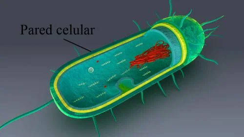
The cell wall isn’t just armor. It’s a defining feature shared by prokaryotes, plant cells, and fungi. This rigid layer sits outside the plasma membrane, acting as the first line of defense.
Its role is surprisingly specific. Without it, cells would collapse under pressure. The wall prevents osmotic shock, keeping the membrane intact when external conditions shift. It locks in shape. No more squishy, undefined blobs.
Dehydration is also kept at bay. In dry environments, this structure holds water where it belongs. It’s not about growth. It’s about survival.
Why do some organisms need this? Because they lack internal scaffolding. Plants rely on turgor pressure against the wall to stand upright. Bacteria use it to resist bursting in hypotonic solutions. Fungi need it to maintain hyphal structure.
It’s rigid. It’s protective. And it’s everywhere those three kingdoms meet.
“The cell wall is a rigid layer outside the plasma membrane, providing structural support and protection against osmotic stress.”
So next time you see a plant standing tall or a fungus spreading across bark, remember: it’s not magic. It’s chemistry. And a very tough outer layer.